A Natural Sciences and Engineering Research Council (NSERC) of Canada Discovery Grant in the amount of $105,000 over five years has been awarded to Dr. Nancy Ford for contrast-enhanced imaging using X-ray and optical techniques. Ford is an assistant professor in the Department of Oral Biological & Medical Sciences and an expert in micro-computed tomography (micro-CT) and in vivo small animal imaging.
Ford’s NSERC-funded study will compare 3D X-ray images from micro-CT and synchrotron studies with optical techniques such as 2D confocal microscopy and 3D optical projection tomography (OPT). The measurements performed on the micro-CT and synchrotron images will provide information about organ structure and function in living animals. The optical techniques will, additionally, provide information about the structure and function of the cells within an organ or tissue. “One of the challenges of using different imaging techniques on a tissue sample is comparing the different data,” Ford explains. “We want to make sure we are comparing apples to apples, so we want to identify contrast agents that are visible using multiple imaging equipment.”

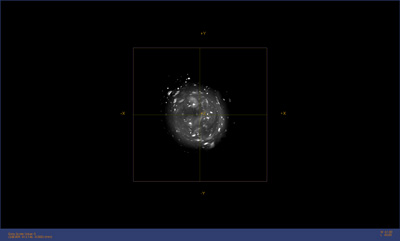
Image from an optical projection tomography scanner, taken
by Dylan Jow, a third-year physics and math major who
undertook UBC’s work–learn program in summer 2016, in
Dr. Nancy Ford’s lab. Jow used maximum intensity projection
to show the brightest pixels in the 3D volume as a 2D image.
The bright dots are quantum dots injected into a block of
agarose, a material similar to gelatin.
Ford’s research lab has a strong foundation in preclinical micro-CT imaging of the lung to develop multi-energy contrast-enhanced micro-CT imaging protocols using iodine and xenon as contrast agents to visualize the blood vessels and airways respectively. She has also developed new imaging techniques on the BioMedical Imaging and Therapy (BMIT) beamline of the Canadian Light Source (CLS) synchrotron facility in Saskatoon, Saskatchewan. These include the first imaging of xenon as a contrast agent using K-edge subtraction (KES) and the first images using respiratory signals from a rodent to trigger image acquisition during specified respiratory phases. Ford’s techniques for micro-CT and on the BMIT beamline will enable in vivo quantitative analysis of structure, physiology and function in rodent organs and tissues.
Ford explains her method: “We will introduce contrast media or staining to visualize the same organ features under different imaging techniques. For in vivo imaging, we traditionally use iodine to visualize the blood vessels in the micro-CT and in KES synchrotron imaging, but this contrast agent is not used for optical imaging. To correlate X-ray and optical techniques, possible contrast agent candidates include gold and silver nanoparticles and CdSe/ZnS quantum dots, which are nanoparticles with a metallic cadmium selenide core and a zinc sulphide shell. We expect all of these materials will be visible in the synchrotron and microcomputed tomography images. Both the silver nanoparticles and quantum dots have been established as optical markers.”
Synchrotron imaging will be performed at the CLS facility using a novel spectral KES technique. For this technique, two images are obtained with different X-ray energies and then the images are subtracted. The energies selected are above and below the K-edge of the nanoparticle contrast material—K-edge is the energy at which X-ray absorption increases dramatically for a given material. Subtracting the images highlights the areas containing the nanoparticles.
The UBC Centre for High-Throughput Phenogenomics (CHTP), where Ford is the director, has two micro-CT scanners—one suitable for imaging live rodents and one for imaging excised organs and tissues. The CHTP also houses an optical projection tomography machine, which produces 3D images of fluorescently labelled tissues, and a white light laser confocal microscope with multiple laser sources.
The novelty of this project includes synchronizing the image acquisition with physiological signals at the BMIT beamline and using a world-wide unique spectral KES technique. The synchrotron will enable very accurate measurements, which will be compared with in vivo micro-CT, ex vivo micro-CT, OPT and microscopy measurements. All together, these data will provide a more complete picture of how an organ functions and identify regions of an organ that are not functioning properly.
To learn more about Dr. Nancy Ford’s research, read the article “In Vivo Imaging—Fascinating Voyage of Discovery” in the fall 2012 issue of Impressions online here >>
To learn more about how the Centre for High-Throughput Phenogenomics can support your scientific objectives, visit chtp.ubc.ca